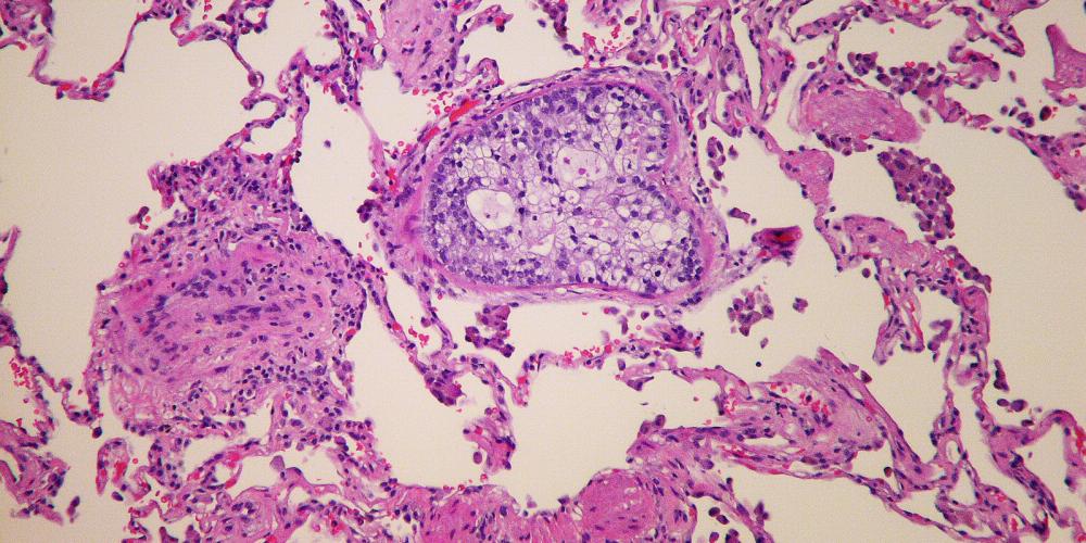
Pancreatic cancer is an aggressive tumour that is very difficult to treat and has low survival rates. Previous studies show that there are two major subtypes of tumour: classical and basal. Basal tumours tend to be more aggressive and are more associated with invasion and metastasis. Much research is being carried out to determine whether the two subtypes respond differently to the most common chemotherapies, which would mean they should each receive targeted treatment.
New research in the Medical and Molecular Oncology Laboratory of Professor Ilse Rooman at VUB is mapping the two types of tumour cells. For her PhD, Ellis Michiels developed a detection method that stains both subtypes in surgical tissue samples. This technique, based on multiple RNA sequences, allows a more detailed view of cancer tissue, and shows that not only do different patients have different subtypes but also that each tumour is composed of both classical and basal tumour cells, and that they can change identity.
“With previous, less sophisticated analyses, patients appeared to belong to one of two groups, but the tumour tissues turn out to be much more complex when one visualises the subtypes,” says Michiels. “The new technique allows quantification of cancer cells of each subtype, which can help to group patients for personalised treatment.” The principle of the detection technique also allows detection of DNA errors, or mutations, in tissue. Michiels: “We were able to detect highly aggressive tumour cells that had assumed the identity of the surrounding tissue and were therefore masked. Our study visually maps the tissue complexity of pancreatic cancer and could be applied in future in clinics as a guide for patient grouping for personalised therapy.”
Reference
Michiels, E. et al. (2023), High-resolution and quantitative spatial analysis reveal intra-ductal phenotypic and functional diversification in pancreatic cancer. J. Pathol. http://doi.org/10.1002/path.6212